Measuring proliferation of T cells, B cells or NK cells can help you asses the immunomodulatory capacity of your drugs or analyze antigen specific reactions. This assay is flow cytometry based and can be easily combined with the analyzes of cytokine production or transcription factor expression.
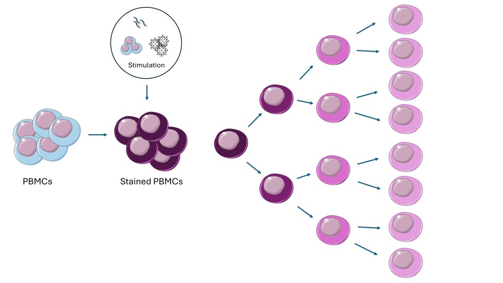
PBMCs stained with a proliferation dye can be stimulated with either other PBMCs (MLR), and antigen, antibody or peptide pool to induce proliferation. The proliferation dye will divide it self evenly over two daughter cells, resulting in a decreasing dye intensity that can by analyzed by flow cytometry.
Antigen specific T cell analysis
The proliferation assay is a very useful tool to determine antigen specific T cells responses, for instance before and after vaccination. Especially when low frequencies are expected and both CD4 and CD8 T cells are investigated. Besides the use of a proliferation dye, there is also the option to use tritiated Thymidine (3HT) to analyze proliferation. This is especially well suited for high throughput screening off vaccine candidates or overall immune modulatory effects. To discuss what would suit your research question best, contact us.
Mixed Lymphocyte reactions
By culturing cells from two different people together an allogeneic immune reaction occurs resulting in T cell activation and proliferation. MLRs can be performed with PBMCs from two donors (two-way MLR) or using monocyte derived Dendritic cells from one donor and T cells from another donor (one-way MLR). An MLR is especially well suited for testing the immune modulating capacity of a drug.
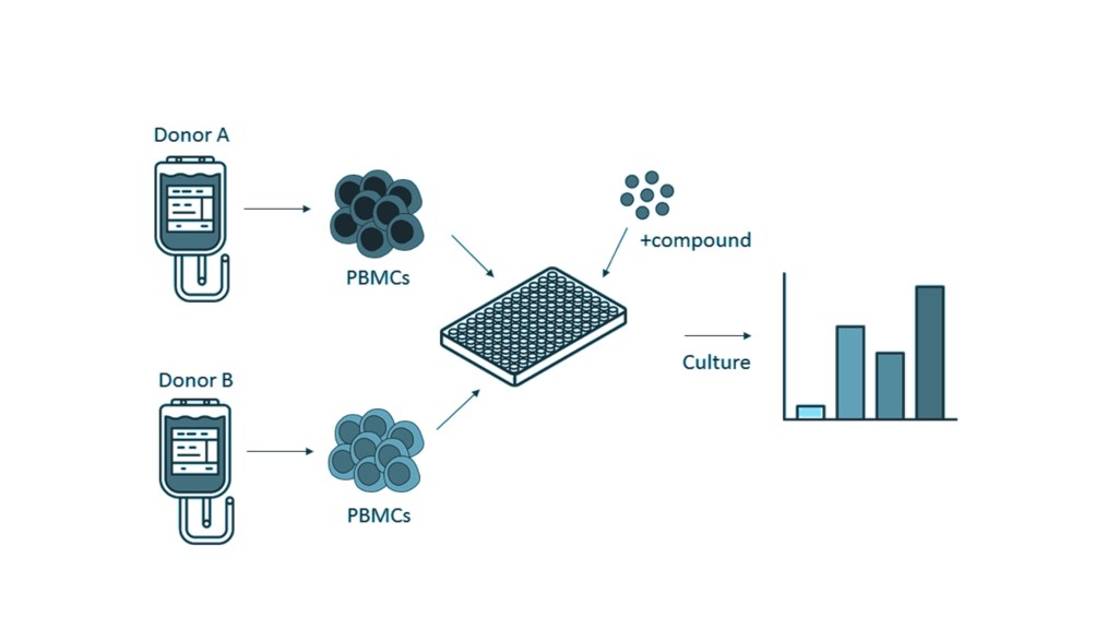
A two way MLR using PBMCs from two different donors to elicit an immune response. The effects of different compounds can be tested by using proliferation or cytokine production as a readout method.
References
Ruben JM, Bontkes HJ, Westers TM, Hooijberg E, Ossenkoppele GJ, van de Loosdrecht AA, de Gruijl TD. In situ loading of skin dendritic cells with apoptotic bleb-derived antigens for the induction of tumor-directed immunity. Oncoimmunology. 2014 Jul 3;3(7):e946360. doi: 10.4161/21624011.2014.946360. PMID: 25610730; PMCID: PMC4292219.
Di Blasi D, Claessen I, Turksma AW, van Beek J, ten Brinke A. Guidelines for analysis of low-frequency antigen-specific T cell results: Dye-based proliferation assay vs 3 H-thymidine incorporation J Immunol Methods 2020;487:112907. doi: 10.1016/j.jim.2020.112907.
ten Brinke A, Marek-Trzonkowska N, Mansilla MJ, Turksma AW, Piekarska K, Iwaszkiewicz-Grześ D, Passerini L, Locafaro G, Puñet-Ortiz J, van Ham SM, Hernandez-Fuentes MP, Martínez-Cáceres EM, Gregori S. Monitoring T-Cell Responses in Translational Studies: Optimization of Dye-Based Proliferation Assay for Evaluation of Antigen-Specific Responses Front Immunol 2017;8:1870. doi: 10.3389/fimmu.2017.01870.
